SOMATOM go.Top
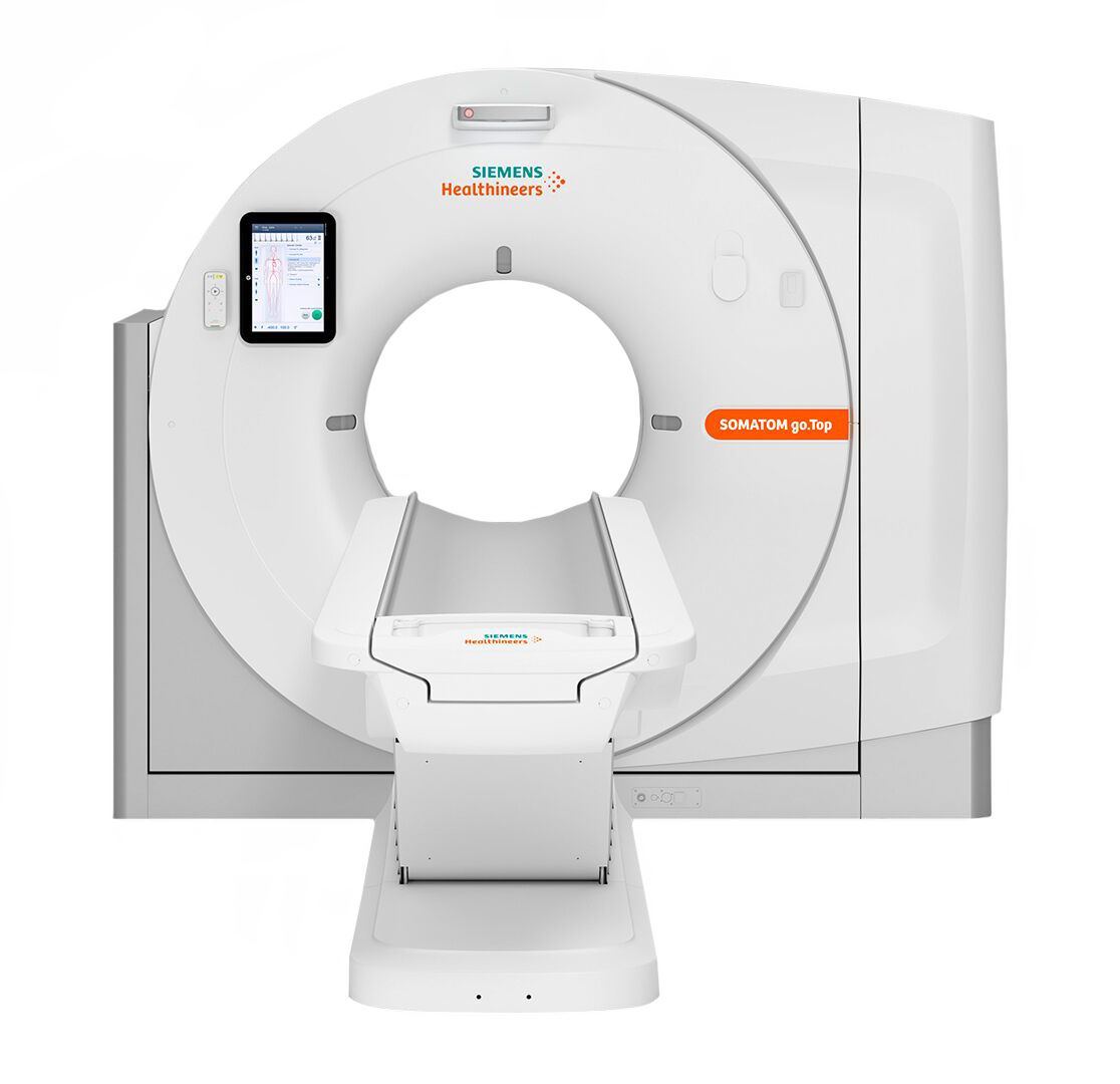

The SOMATOM go.Top system provides a complete set of intuitive solutions aimed at optimizing your workflow not only at the scanner, but also beyond it. Today, these functions are available both for routine scanning and for scanning in advanced clinical areas. By reducing the number of repetitive workflow steps, GO technologies help standardize and simplify all processes in the department – from setting up patient parameters to distributing, archiving and reading images. Therefore, you can work more efficiently and focus on your patients – these two key factors ensure successful business.
Features and Benefits
The main element of optimizing efficiency and improving patient comfort is a completely new approach to working with the scanner. The SOMATOM go.Top system, created on the basis of a new mobile workflow, offers a number of innovative solutions. A tablet, a remote control, a camera, a built-in mount for a contrast agent injector* and a new workplace design provide an unprecedented level of flexibility and mobility during daily CT procedures.
CARE Dose 4D
CARE Dose4D primarily uses automatic adjustment of the dose level depending on the patient’s body size based on the signal attenuation values obtained from a standard (one-time) topogram along the patient’s z axis. In addition, CARE Dose4D uses tube current adaptation based on the actual attenuation of the X-ray beam measured around the patient’s body. CARE Filter: A specially designed butterfly filter for X-ray irradiation mounted on a collimator.
CARE kV, 10 kV Steps, CARE Child
The CARE kV option automatically sets the tube voltage depending on the characteristics of each patient and clinical indications. Due to the optimal voltage level in each case, the CARE kV option allows you to keep the dose low, which makes it ideal for use in pediatric imaging. This further simplifies the process by equalizing the tube current with the selected voltage value. Our unique 10 kV Steps feature also helps to adapt the voltage to your patient’s parameters. This function adjusts the level at intervals of 10 kV for, which provides a lower dose and high contrast resolution. The CARE Child function offers a number of targeted solutions to minimize radiation exposure while maintaining high quality diagnostic images. Pediatric protocols automatically set the low voltage of the tube, in most cases this value is 70 kV, while the CARE Dose4D function optimizes the dose distribution and offers special modulation curves. X-ray irradiation is set depending on the weight and age of the child (and an adult of small stature and weight), which significantly reduces the effective dose for the patient.
CARE Topo
The topogram is formed in real time; it is possible to interrupt the formation manually after obtaining the desired anatomy.
CARE Bolus
Operating mode for collecting data obtained by contrast expansion. The goal is to optimally use a bolus of contrast agent in the “plateau” phase in the target organ. This option has been specially adapted to the increased speed and time requirements due to the possibility of using multiple rows and faster rotation. Contrast expansion is monitored by monitoring scanning in a user-defined area while observing the trigger threshold. Spiral scanning starts as soon as the expansion reaches a predetermined threshold value.
CARE Profile
Visualization of dose distribution by topogram before scanning Topogram: Scanning perspectives: anterior-posterior view (ap), posterior-anterior view (pa), lateral view (lat);
Image reconstruction and storage: 512 x 512 reconstruction matrix; reconstruction fields from 5 cm to 70 cm (with HD FoV Pro) using scaling of the source data with the possibility of freely choosing the image center before scanning (perspective) or after it (retrospectively). Storing images and raw patient data.
HD FoV Pro
This technology is designed to visualize parts of the human body and skin located outside the standard 50-cm scanning field of view to the size of the gantry hole, based on algorithmic addition of missing sensor data outside the standard 50-cm scanning field of view. The image quality for an area outside the standard 50-cm scanning field of view does not match the image quality of the area inside such a field of view. Depending on the settings and the scanned area of the patient’s body, image artifacts may appear.
DynSerio Scan
DynSerio Scan provides dynamic scanning using sensor width. Data is collected at different points in time from one anatomical point, while the table on which the patient is located remains stationary.
Workstream4D
The WorkStream 4D function further expands the workflow by directly generating sagittal, coronal, oblique or double oblique reconstructed images directly from the original CT data within the CT protocol. Unlike other automated multi-plane Reformation (MPR) solutions, WorkStream 4D does not require reconstruction of thin slice data before creating reformatted images. This provides time savings compared to alternative MPR methods. In addition, WorkStream 4D allows the user to create oblique and double oblique reformations in the form of MPR or MIP images, which greatly facilitates the workflow for both routine and CTA analysis compared to alternative methods.
IVR (interlaced volume reconstruction)
Use measurement data as efficiently as possible with interlaced volumetric reconstruction (IVR). The ability to extract the maximum amount of diagnostic information from measurement data The ability to improve spatial sampling in the z direction, regardless of the step value, Evaluation of the smallest structures, such as injuries or fractures.
X-CARE
X-CARE is a partial scanning function designed to reduce direct X-ray exposure to dose-sensitive areas of the body, for example, the lens of the eye.
Adaptive signal amplification
The adaptive signal amplification function amplifies low signals when they are strongly suppressed, for example, when imaging obese patients or patients with metal implants. This reduces artifacts in the form of stripes, ensuring that the correct H values are maintained without compromising spatial resolution. Signal quality analysis and integration of information from neighboring sensor elements in low-signal areas can significantly reduce image noise.
DoseMAP
DoseMAP is a dosage management program in Siemens tomographs that provides transparency of dose values and allows you to assess the dosage situation. Setting alarm alerts when the dose is exceeded increases safety. The DoseMAP program has three components for complete and comprehensive dose management: Reporting, Analysis and Protection.
Similar products